Home » Without Label » Upper Leg Tendon Anatomy / Muscles of the Leg (Human) / This is why you have to indicate which biceps you are taking about when discussing one or other of these muscles.
Upper Leg Tendon Anatomy / Muscles of the Leg (Human) / This is why you have to indicate which biceps you are taking about when discussing one or other of these muscles.
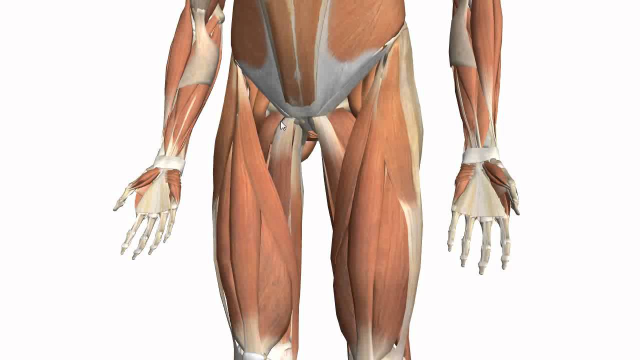
Upper Leg Tendon Anatomy / Muscles of the Leg (Human) / This is why you have to indicate which biceps you are taking about when discussing one or other of these muscles.. Lie prone on a hamstring curl machine. The only muscle of the quadriceps to cross both the hip and knee joints. The knee joint is the junction of the thigh and leg. Ebraheim's educational animated video describes muscle anatomy of the thigh. The legs are the lower limbs of the human body that provide support and stability in addition to allowing movement.
Tendons are thick bands of tissue that connect muscles to bone. The thigh has three sets of strong muscles: Upper leg tendon anatomy : Upper leg muscle pain is a very hard pain affect the leg pain as a whole. Lateral (fibular) collateral ligament (fcl) upper part middle part lower part popliteus tendon (pt) upper part i.
Before Fight Start Hand Boxer Ring Stock Photo 340800278 ... from thumb9.shutterstock.com This tendon helps your leg bend when you raise your knee. Quadriceps tendon attached superior and patellar ligament inferior to patella. To download this image, create an account. The gastrocnemius is the bulging muscle that's most visible. Learn about the muscles, tendons, bones, and ligaments that comprise the knee joint anatomy. When we knowing the causes of the upper leg muscle pain it will be simple to treat and relieve the pain. It also arises from the base of the greater trochanter and the linea aspera, the supracondylar ridge, and the lateral intermuscular septum. The tendons for these muscles begin at your ischial tuberosity, or ischium (the bony bump under each buttock), and attach on the outer edges of your shinbones (tibia and fibula) just below the back of your knee.
The knee joint is the junction of the thigh and leg.
Other muscles of the anterior (front) thigh include the pectineus, sartorius,. The legs are the lower limbs of the human body that provide support and stability in addition to allowing movement. Upper leg tendon anatomy : It allows your foot to flex as you walk or run. Lie prone on a hamstring curl machine. The quadriceps tendon attaches the quadriceps muscles to the patella. The posterior upper leg muscles provide your knees with mobility (extension, flexion and rotation) and strength. It's flat and thick, rising from the bones of the tibia and. If you feel it you need to take care of the causes of this hard pain. There are a number of bones, muscles, and tendons in the area. Meanwhile, the vastus lateralis is on the side of the thigh, while the vastus intermedius is hidden below the rectus femoris(5). The rectus femoris is located in the center of the thigh, while the vastus medialis is in the middle of the said body part. Tendons are thick bands of tissue that connect muscles to bone.
Notice the upper leg has a biceps muscle just like the upper arm does. Use the mouse scroll wheel to move the images up and down alternatively use the tiny arrows (>>) on both side of the image to move the images. A muscle strain (muscle pull or tear) is a common injury, particularly among people who participate in sports. Anatomy the four quadriceps muscles meet just above the kneecap (patella) to form the quadriceps tendon. The soleus muscle lies underneath the gastrocnemius.
BIOL 160: Human Anatomy and Physiology | Human anatomy ... from i.pinimg.com The leg anatomy includes the quads, hams, glutes, hip flexors, adductors & abductors. It's flat and thick, rising from the bones of the tibia and. Anatomy the four quadriceps muscles meet just above the kneecap (patella) to form the quadriceps tendon. The tendons for these muscles begin at your ischial tuberosity, or ischium (the bony bump under each buttock), and attach on the outer edges of your shinbones (tibia and fibula) just below the back of your knee. Upper leg anatomy and function. Pin on upper leg muscle anatomy from i.pinimg.com the human leg, in the general word sense, is the entire lower limb of the human body, including the foot, thigh and even the hip or gluteal region. The patellar tendon is an essential tendon in the leg that enables the muscles to. Its muscle belly is on the back aspect of the upper arm.
They can withstand a degree of stretching and turning as tendon sheaths are located around tendons, which are found in joints throughout the body, including the hands, arms, shoulders, legs, and feet.the human leg, in the general word sense, is the entire lower limb of the human body.
This tendon helps your leg bend when you raise your knee. The word sartorius is derived from the latin word sartor, which translates to patcher, or tailor, due to the way the individual will position their leg while working. Tendons are thick bands of tissue that connect muscles to bone. The patellar tendon is an essential tendon in the leg that enables the muscles to. On the medial edge of the posterior thigh is the gracilis muscle. Ebraheim's educational animated video describes muscle anatomy of the thigh. Upper leg tendon anatomy this mri wrist coronal cross sectional anatomy tool is absolutely free to use. The tendons for these muscles begin at your ischial tuberosity, or ischium (the bony bump under each buttock), and attach on the outer edges of your shinbones (tibia and fibula) just below the back of your knee. Upper leg anatomy and function the upper leg is often called the thigh. See more ideas about muscle anatomy, leg muscles anatomy, anatomy. Meanwhile, the vastus lateralis is on the side of the thigh, while the vastus intermedius is hidden below the rectus femoris(5). Muscles of the arm at la. Upper leg anatomy and function.
It serves to attach the plantaris, gastrocnemius (calf) and soleus muscles to the calcaneus (heel) bone. Anatomy the four quadriceps muscles meet just above the kneecap (patella) to form the quadriceps tendon. Upper leg tendon anatomy : The leg anatomy includes the quads, hams, glutes, hip flexors, adductors & abductors. Lateral (fibular) collateral ligament (fcl) upper part middle part lower part popliteus tendon (pt) upper part i.
Muscles of the Thigh Part 2 - Medial Compartment - Anatomy ... from i.ytimg.com If you feel it you need to take care of the causes of this hard pain. Lateral (fibular) collateral ligament (fcl) upper part middle part lower part popliteus tendon (pt) upper part i. The sartorius is the longest muscle in the body, spanning both the hip and the knee joints. The only muscle of the quadriceps to cross both the hip and knee joints. The lower leg lies between the knee and ankle and works with the upper leg and foot to help perform the key functions of the leg. The legs include the upper leg, knee, lower leg, ankle, and. The hamstring muscles in the back of the thigh, the quadriceps muscles in the front, and the adductor muscles on the inside. #muscle and tendon pain in legs #muscles and tendons of the leg and foot #muscles and tendons of the lower leg #muscles ligaments and tendons of the lower leg #muscles tendons and ligaments of the upper leg
The posterior upper leg muscles provide your knees with mobility (extension, flexion and rotation) and strength.
The knee joint is the junction of the thigh and leg. The patella is attached to the shinbone (tibia) by the patellar tendon. Muscles of the arm at la. Meanwhile, the vastus lateralis is on the side of the thigh, while the vastus intermedius is hidden below the rectus femoris(5). This tendon helps your leg bend when you raise your knee. It allows your foot to flex as you walk or run. The lower leg lies between the knee and ankle and works with the upper leg and foot to help perform the key functions of the leg. Lie prone on a hamstring curl machine. This is why you have to indicate which biceps you are taking about when discussing one or other of these muscles. Upper leg tendon anatomy : The legs include the upper leg, knee, lower leg, ankle, and. Learn about the muscles, tendons, bones, and ligaments that comprise the knee joint anatomy. The gastrocnemius is the bulging muscle that's most visible.